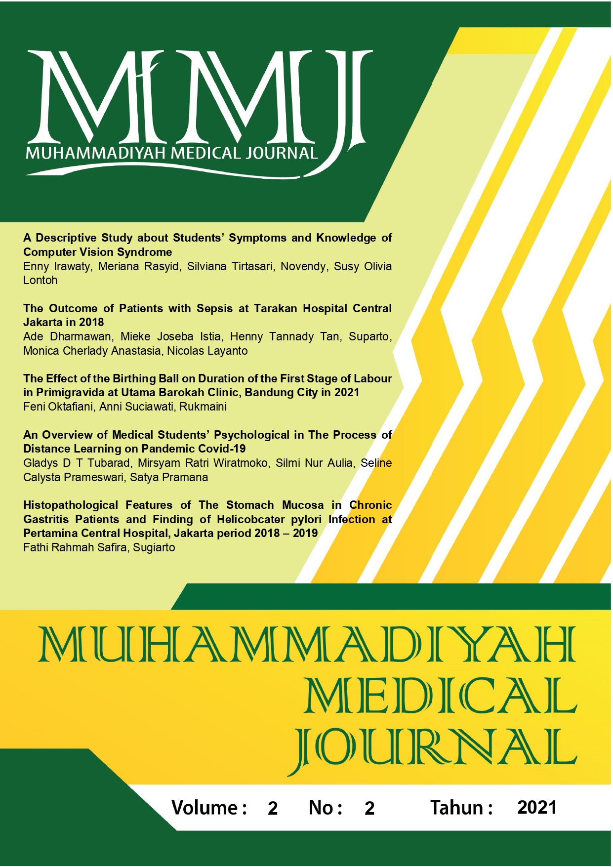
Histopathological Features of the Gastric Mucosa in Patients with Chronic Gastritis and Helicobacter pylori Infection at Pertamina Central Hospital Jakarta
DOI:
https://doi.org/10.24853/mmj.2.2.70-74Keywords:
dysplasia, gastric mucosal atrophy, histopathology of chronic gastritis, Helicobacter pylori, intestinal metaplasiaAbstract
Background: Chronic gastritis is a chronic inflammation of the gastric mucosa, accompanied by changes in mucosal histology with or without Helicobacter pylori infection. Changes in the gastric mucosa include gastric mucosal atrophy, intestinal metaplasia, and epithelial dysplasia. Purposes: This study aims to determine the microscopic appearance of the mucosa in chronic gastritis patients based on standard histopathological criteria, which include gland atrophy, intestinal metaplasia, dysplasia with or without Helicobacter pylori infection at Pertamina Central Hospital Jakarta period 2018 - 2019. Methods: This retrospective study was conducted in a cross-sectional study from March 15 to March 25, 2020. Results: This study reported 303 cases of active chronic gastritis (38.4%) out of 790 total samples that met the inclusion criteria. Microscopic changes of the mucosa were found in the form of atrophy of the mucous glands in 254 cases (32.2%), intestinal metaplasia in 25 cases (3.2%), and epithelial dysplasia cases in 23 cases (2.9%). Conclusion: the proportion of active chronic gastritis patients found in this study was helicobacter pylori infection is more dominantly found in patients with active chronic gastritis than in non-active chronic gastritis. The description of atrophic glands in chronic gastritis patients was more dominant than parameters based on intestinal metaplasia and epithelial dysplasia. It was a finding of intestinal metaplasia compared to all cases showing risk factors that require further clinical observation (follow-up) to detect potential malignancies earlier so that it is necessary to do more preventive action.References
Marcus AJ. Chronic Gastritis. Medscape. 2019;(Chronic Gastritis).
Hirlan. Gastritis. In: Ilmu Penyakit Dalam Jilid 2 Ed VI. 2014. p. 1770.
Brooks GF. Mikrobiologi Kedok teran Jawetz, Melnick, & Adelberg, Ed.25. 2010.
Kumar V, Abbas AK, Aster J. Buku Ajar Patologi Robbins Edisi 9. 9th ed. Elsevier Saunders; 2015.
Vakil N. Overview of Gastritis. MSD Manual. 2020.
Zhang C. Helicobacter pylori Infection, Glandular Atrophy, and Intestinal Metaplasia in Superficial Gastritis, Gastric Erosion, Erosive Gastritis, Gastric Ulcer, and Early Gastric Cancer. World J Gastroenterol. 2005;791–6.
Emory TS. Atlas of Gastrointestinal Endoscopy and Endoscopic Biop-sies. 2000.
Jayanthi C, M L, Kumar GR, Kumar SA. Histopathological analysis of chronic gastritis and correlation of pathological features with helicobac-ter pylori. Indian J Pathol Oncol. 2017;4(4):495–500.
Watari J, Chen N, Amenta PS, Fukui H, Oshima T, Tomita T, et al. Helicobacter pylori associated chronic gastritis, clinical syndromes, precancerous lesions, and patho-genesis of gastric cancer develop ment. World J Gastroenterol. 2014;20(18):5461–73.
Garg B, Sandhu V, Sood N, Sood A, Malhotra V. Histopathological analysis of chronic gastritis and correlation of pathological features with each other and with endoscopic findings. Polish J Pathol. 2012;63 (3):172–8.
Rugge M, Pennelli G, Fassan M, Pilozzi E, Ingravallo G, Russo V, et al. Gastritis : The Histology Report. Dig Liver Dis. 2011;373–84.
Dîrnu R, Secureanu FA, Neamţu C, Totolici BD, Pop OT, Mitruţ P, et al. Chronic gastritis with intestinal metaplasia: Clinico-statistical, histo-logical and immunohistochemical study. Rom J Morphol Embryol. 2012;53(2):293–7.
Saxena M. Prevalence of Intestinal Metaplasia and Dysplasia in Infectious and Non-infectious Chronic Gastritis. Int J Res Med Sci. 2020;6.
Lim JH, Kim N, Lee HS, Choe G, Jo SY, Chon I, et al. Correlation between endoscopic and histological diagnoses of gastric intestinal metaplasia. Gut Liver. 2013;7(1): 41–50.
Sung JK. Diagnosis and Manage ment of Gastric Dysplasia. Korean J Intern Med. 2016;31(2): 201–9.
Downloads
Published
Issue
Section
License
Authors who publish in the Muhammadiyah Medical Journal agree to the following terms:
- Authors retain copyright and grant Muhammadiyah Medical Journal right of first publication with the work simultaneously licensed under a Creative Commons Attribution Licence that allows others to adapt (remix, transform, and build) upon the work non-commercially with an acknowledgement of the work's authorship and initial publication in Muhammadiyah Medical Journal.
- Authors are permitted to share (copy and redistribute) the journal's published version of the work non-commercially (e.g., post it to an institutional repository or publish it in a book), with an acknowledgement of its initial publication in Muhammadiyah Medical Journal.


