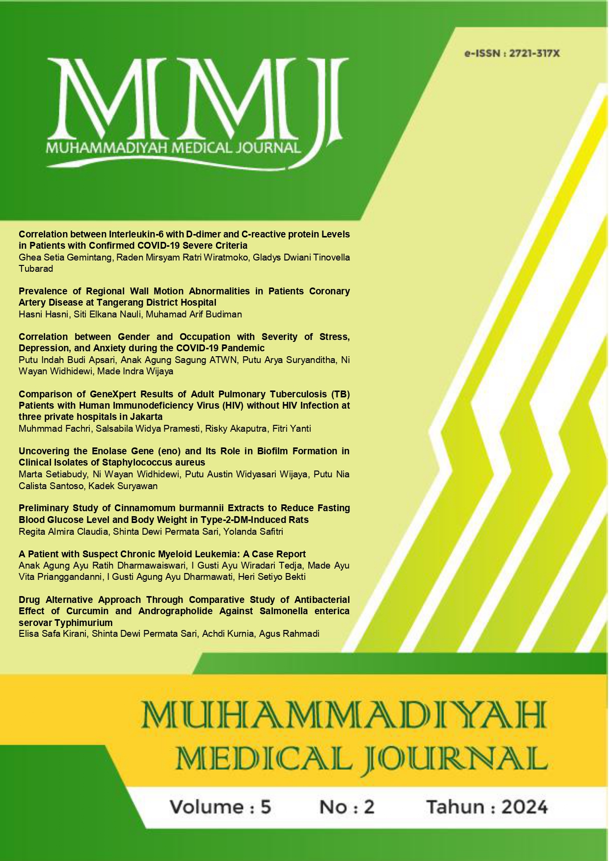
Prevalence of Regional Wall Motion Abnormalities in Patients Coronary Artery Disease at Tangerang District Hospital
DOI:
https://doi.org/10.24853/mmj.5.2.76-82Keywords:
CAD, RWMA, WMSI, bull's-eye plot parameterAbstract
Background: Coronary artery disease (CAD) is when the coronary blood vessels cannot supply blood to the heart due to a pile of atherosclerotic plaque in the coronary arteries, so blood flow to the myocardium is disrupted. This blood flow disturbance will cause myocardial contractile dysfunction and Regional Wall Motion Abnormalities (RWMA). Purposes: This study aims to determine the prevalence of Regional Wall Motion Abnormalities in CAD patients based on a single-center study at Tangerang District Hospital in January - May 2024. Methods: Our study used a descriptive approach in 130 CAD patients grouped based on gender, age, hypertension, and diabetes mellitus. In addition, the determination of RWMA severity cases in CAD was measured by only the Wall Motion Score Index (WMSI) and Wall Motion Score Index (WMSI) followed by the Bull's-eye plot parameters. Results: The prevalence of CAD patients with RWMA using the WMSI method was found to be 41 patients (32%) who were predominantly male (76%) and occurred at the mean of age 61,14 years, 27% hypertension, and 17% diabetes mellitus. Conclusion: WMSI parameters followed by the Bull's-eye plot could identify 14.6% more RWMA cases than only WMSI.References
Jeetley P, Khattar RS, Senior R. Coronary Artery Disease: Assessing Regional Wall Motion. In: Echocardiography, Second Edition. 2018.
Johnson NB lai., Hayes LD, Brown K, Hoo EC, Ethier KA. CDC National Health Report: leading causes of morbidity and mortality and associated behavioral risk and protective factors--United States, 2005-2013. MMWR Surveill Summ. 2014;63.
Brown JC, Gerhardt TE KE. Risk Factors For Coronary Artery Disease. [Updated 2022 Jun 5]. StatPearls [Internet] Treasure Isl StatPearls Publ 2022. 2022;
Kementerian Kesehatan RI Badan Penelitian dan Pengembangan. RISKESDAS 2018. Kementerian Kesehat Republik Indones. 2018;
Espersen C, Modin D, Platz E, Jensen GB, Schnohr P, Prescott E, et al. Global and regional wall motion abnormalities and incident heart failure in the general population. Int J Cardiol. 2022;357.
Playford D, Stewart S, Harris SA, Chan YK, Strange G. Pattern and Prognostic Impact of Regional Wall Motion Abnormalities in 255 697 Men and 236 641 Women Investigated with Echocardiography. J Am Heart Assoc. 2023;12(22).
Khera A V., Kathiresan S. Genetics of coronary artery disease: Discovery, biology and clinical translation. Vol. 18, Nature Reviews Genetics. 2017.
Widiantoro S, Hamed O. Korelasi Antara Skor Indeks Gerakan Dinding Jantung Dengan Fraksi Ejeksi Pada Penyakit Jantung Koroner. ARKAVI; 2016. p. 66–76.
McGuire S, Horton EJ, Renshaw D, Chan K, Jimenez A, Maddock H, et al. Cardiac stunning during haemodialysis: the therapeutic effect of intra-dialytic exercise. Clin Kidney J. 2021;14(5).
Radwan H, Hussein E. Value of global longitudinal strain by two dimensional speckle tracking echocardiography in predicting coronary artery disease severity. Egypt Hear J. 2017;69(2).
Wierzbowska-Drabik K, Picano E, Simiera M, Plewka M, Krȩcki R, Peruga JZ, et al. A head-to-head comparison of wall motion score index, force, strain, and ejection fraction for the prediction of SYNTAX and Gensini coronary scores by dobutamine stress echocardiography. Kardiol Pol. 2020;78(78).
Abdel Mawla TS, Abdel Wanees WS, Abdel Fattah EM, El khashab KA, Momtaz OM. Diagnostic accuracy of global longitudinal strain in prediction of severity and extent of coronary artery stenosis in patients with acute coronary syndrome. Acta Cardiol. 2023;78(1).
Gaibazzi N, Bergamaschi L, Pizzi C, Tuttolomondo D. Resting global longitudinal strain and stress echocardiography to detect coronary artery disease burden. Eur Heart J Cardiovasc Imaging. 2023;24(5).
Alaika O, Jamai S, Doghmi N, Cherti M. Diagnostic accuracy of global longitudinal strain for detecting significant coronary artery disease in diabetic patients without regional wall motion abnormality. J Saudi Hear Assoc. 2020;32(3).
Tampubolon LF, Ginting A, Saragi Turnip FE. Gambaran Faktor yang Mempengaruhi Kejadian Penyakit Jantung Koroner (PJK) di Pusat Jantung Terpadu (PJT). J Ilm Permas J Ilm STIKES Kendal. 2023;13(3).
Dahliah, Hidayati PH, Wisudawan, Wahab MI, Humairah AR. Analisis Faktor-Faktor Risiko Kejadian Penyakit Jantung Koroner. 2024;6(4).
Woods MD, Hatfield J, Hammonds K, Exaire J, Mixon TA, Nguyen V, et al. Regional wall motion abnormalities in transthoracic echocardiography in patients with significant coronary artery disease and coronary collateral circulation in adults. Cardiovasc Revascularization Med. 2024;(May).
Sarker MHR, Moriyama M, Rashid HU, Chisti MJ, Rahman MM, Das SK, et al. Community-based screening to determine the prevalence, health and nutritional status of patients with CKD in rural and peri-urban Bangladesh. Ther Adv Chronic Dis. 2021;12.
Pate A, Emsley R, Sperrin M, Martin GP, van Staa T. Impact of sample size on the stability of risk scores from clinical prediction models: a case study in cardiovascular disease. Diagnostic Progn Res. 2020;4(1).
Rosen-Wetterholm E, Cavefors O, Redfors B, Ricksten SE, Omerovic E, Polte CL, et al. RWMAs in critically ill patients with non-obstructed coronary arteries. Acta Anaesthesiol Scand. 2023;67(6).
Tserioti E, Chana H, Salmasi AM. Hypertensive patients are more likely to develop coronary artery disease on computed tomography coronary angiography. Eur J Prev Cardiol. 2023;30(Supplement_1).
Shahriar MS, Haque T, Rahman AU, Rahman H, Shaila U. Strain Analysis Using Speckle Tracking Echocardiography for Detection of Coronary Artery Disease in Stable Angina Patients with No Regional Wall Motion Abnormality at Rest. Sch J Appl Med Sci. 2022;10(10).
Downloads
Published
Issue
Section
License
Authors who publish in the Muhammadiyah Medical Journal agree to the following terms:
- Authors retain copyright and grant Muhammadiyah Medical Journal right of first publication with the work simultaneously licensed under a Creative Commons Attribution Licence that allows others to adapt (remix, transform, and build) upon the work non-commercially with an acknowledgement of the work's authorship and initial publication in Muhammadiyah Medical Journal.
- Authors are permitted to share (copy and redistribute) the journal's published version of the work non-commercially (e.g., post it to an institutional repository or publish it in a book), with an acknowledgement of its initial publication in Muhammadiyah Medical Journal.


